

 РУ
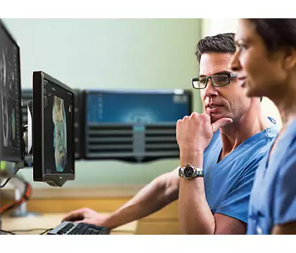
QLAB Cardiology Analysis Software Package
Specialized software for quantitative analysis of the heart and blood vessels
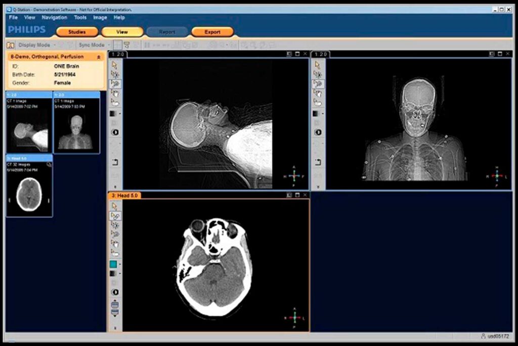
QLAB 13 is designed to provide the information needed to make decisions that will ensure high-quality patient care and more efficient staff work. Improve department work with the ability to query and view data both on the system itself and on a separate workstation.
Contact usGallery


Description
Cardiac 3D Quantification (3DQ)
Cardiac 3D Quantification (3DQ) allows you to view, display, and slice 3D images. From the measured distances and areas in 2D MPR images, you can calculate the LV volume in two planes, ejection fraction (EF), and LV mass. Manipulating 2D planes allows you to more accurately calculate EF in two 2D planes without geometric projection distortions.
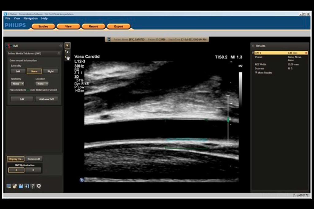
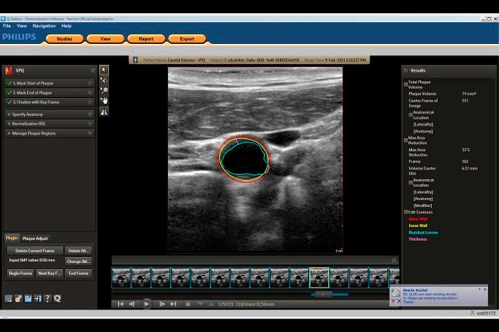
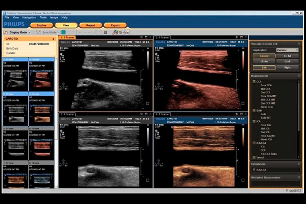
Intima-media thickness (IMT) assessment
Automatic measurement of intima-media thickness in carotid arteries. Fast and easy acquisition of IMT data.
Mitral Valve Navigator (MVNᴬᴵ)
A simple tool for step-by-step creation of a complete list of all necessary measurements and calculations for the mitral valve. Requires acquisition of a volume image in Live 3D TEE mode. Creates an easily interpretable model in just eight steps. The most commonly used orifice and leaflet measurements can be performed in just four steps.
Advanced 3D Cardiac Quantitative Analysis (3DQA)
Measuring LV volumes, stroke volume (SV) and ejection fraction (EF) from 3D data using semi-automated 3D boundary detection. Evaluates the timing of each of 17 minimal regional volumes and determines a 3D synchrony index for all segments or a user-selected group of segments. The semi-automated quantification function allows for the measurement of overall ejection fraction from 3D data without geometric assumptions. It also provides simultaneous access to multiple information for heart failure assessment.
Regions of Interest (ROI) Analysis
This module allows you to obtain echo-contrast and color images and helps extract acoustic data from them.
Strain Quantitative (SQ)
This quantification function generates functional data from tissue color mapping and Doppler velocity data and helps calculate displacement, strain, and strain rate.
Automated 2D Cardiac Quantification (a2DQᴬᴵ)
Automatic LV contouring. Fast and reliable calculation of ejection fraction and volumes in 2D mode. It is possible to choose between the Simpson method / TMAD method of measurement in two planes. a2DQᴬᴵ is ideal for any ultrasound diagnostic room – it allows you to calculate the ejection fraction of any patient with one click, without manual contouring. TMAD method provides a proven indicator that correlates with ejection fraction, which is especially useful for patients with a poor ultrasound window.
Automated Cardiac Motion Quantification (aCMQᴬᴵ)
Based on speckle tracking in 2D images, this function automatically builds a region of interest based on the selected anatomical projection and measures global and regional myocardial function. It then generates a report in the form of a table, a 17-segment bull’s eye diagram, and various curves. This function also calculates the ejection fraction (EF), systolic volume (ESV), and diastolic volume (EDV) for the left ventricle. This is a quick and easy way to simultaneously measure the ejection fraction and global longitudinal strain from the acquired images.
Quantitative Analysis of Cardiac Mechanics under Stress (CMQ Stress)
A method based on speckle tracking technology in 2D images to assess global and regional cardiac function at rest and under maximum load. A fast and user-friendly interface designed specifically for stress echographic studies.
Accurate and fast calculation of ejection fraction
Ejection fraction is assessed in almost every ultrasound examination of the heart. It is one of the most important echocardiographic parameters used to predict left ventricular function and deterioration of the clinical picture of the disease. In addition, it is one of the most important factors taken into account when planning surgical intervention. QLAB software package for calculating ejection fraction (EF); Automated Cardiac 2D Quantificationᴬᴵ — a2DQᴬᴵ (automated cardiac 2D quantification function), Automated Cardiac Motion Quantificationᴬᴵ — aCMQᴬᴵ (automated cardiac motion quantification), Cardiac 3D Quantification — 3DQ (quantitative analysis of cardiac 3D images), Advanced Cardiac 3D Quantification — 3DQ Advanced (advanced cardiac 3D quantification)
Greater confidence in ischemia diagnosis
Stress echo can become a key tool for ischemia diagnosis. However, if it fails to provide accurate results, physicians lose time needed for diagnosis and treatment. Philips has developed technologies that simplify the acquisition of images needed to provide confident results. PureWave technology delivers exceptional image quality in the 1–5 MHz frequency range, with excellent penetration depth and minimal artifacts, even in technically challenging patients with poor ultrasound windows. The X5-1 xMATRIX transducer with iRotate enables the entire stress echo protocol to be performed from a single window without moving the transducer.
Treatment of patients with heart failure
Cardiac resynchronization therapy can improve survival rates in patients with heart failure. However, existing methods for predicting a positive response are not always accurate, resulting in expensive treatments being used ineffectively. However, any new assessment method must be easily integrated into standard ultrasound examinations. The QLAB package offers comprehensive 2D and 3D analysis tools for assessing patients with heart failure and includes IMT (Intima-Media Thickness Measurement), Automated Cardiac Motion Quantification — aCMQᴬᴵ (Automated Cardiac Motion Quantification), Advanced Cardiac 3D Quantification — 3DQ Advanced (Advanced Cardiac 3D Quantification).
Proven Quantitative Analysis Tool
QLAB is for you if you are looking for specialized quantitative analysis tools to simplify your studies, improve their reproducibility, and achieve new levels of clinical insight; if you need to view, manipulate, and measure 3D data sets with detailed clinical reporting; if you need advanced 2D and 3D quantification, as well as quantification of color Doppler and contrast-enhanced images; if you need stand-alone viewing, acquisition, and quantification of 2D, color Doppler, and 3D images with the ability to quickly and easily create BMP, TIF, JPG, MOV, WMV, and AVI files.