
 РУ




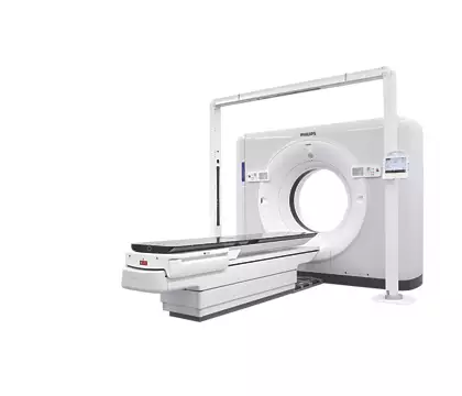
The Philips CT Big Bore RT is a CT simulator that improves planning accuracy, delivering effective treatment for patients, confidence in outcomes, and streamlined workflow for clinicians.
CT Big Bore offers advanced tools to facilitate accurate efficient patient marking and simulation workflow. Featuring 60 cm true scan FOV for full anatomic visualization. It provides spatial positioning accuracy of less than 2mm between the imaging plane and the laser marking plane for confidence in patient marking. This meets the AAPM TG 66.
[/col_inner_3] [col_inner_3 span=”6″ span__sm=”12″]While excellent image quality is the foundation for accurate contouring and treatment plans, radiation oncology demands more. The CT Big Bore offers superb imaging and simulation solutions that integrate with your department workflow for the accuracy and efficiency cancer care requires.
[/col_inner_3] [col_inner_3 span=”6″ span__sm=”12″]The Pulmonary Toolkit for Oncology provides a comprehensive set of tools for evaluating tumor motion and aids in making clinical decisions
[/col_inner_3] [col_inner_3 span=”6″ span__sm=”12″]Efficiently move from scan to plan with virtual simulation and patient marking integrated at the scanner. The Philips TumorLOC application offers an accurate and efficient workflow for patient marking and virtual simulation directly from the scanner console. Tools assist with isocenter localization, generating maximum, minimum and average intensity projections, contouring target volumes and critical structures, beam placement, and machine characterizations for routine and respiratory-correlated studies.
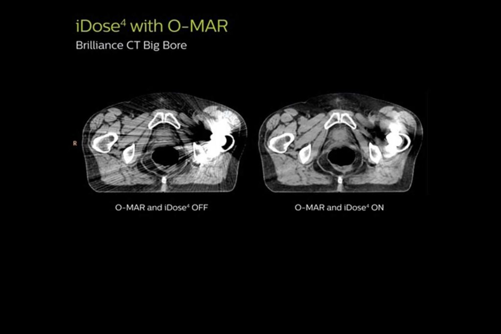
[/col_inner_3] [col_inner_3 span=”6″ span__sm=”12″]Philips provides two leading technologies that can improve image quality. iDose4* improves image quality* through artifact prevention and increased spatial resolution at low dose. O-MAR reduces artifacts caused by large orthopedic implants. Together they product high image quality with reduced artifacts.
[/col_inner_3] [col_inner_3 span=”6″ span__sm=”12″]iPatient provides patient-centered imaging – including imaging for simulation and treatment planning – providing consistent image quality from scan to scan. iPatient features a simple user interface to manage image quality, dose, and speed of acquisition. Drive confidence and consistency with dedicated ExamCards for oncology as well as diagnostic CT procedures.
[/col_inner_3] [/row_inner_3]