

 РУ

Q-Station package in cardiology
Philips Cardiovascular Ultrasound
Q-Station software helps you optimize your workflow and perform specialized quantitative analysis of your Philips ultrasound data. Q-Station includes a wide range of capabilities that allow you to perform any task autonomously.
Contact usGallery


Description
Built-in Q-Assistant and Q-Setups tools simplify setup
The Q-Assistant app lets you select parameters, media, and connection and backup methods. The Q-Setups app lets you set up measurement and analysis packages and includes tools for mapping ultrasound measurements from other manufacturers.
A suite of features to optimize and customize your workflow
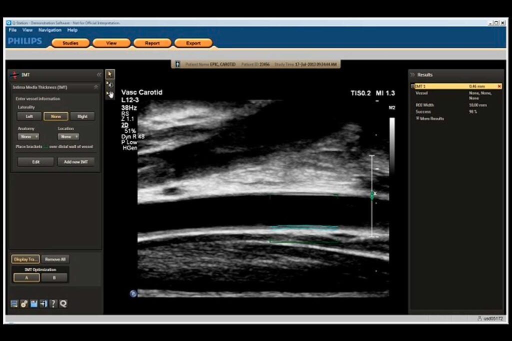
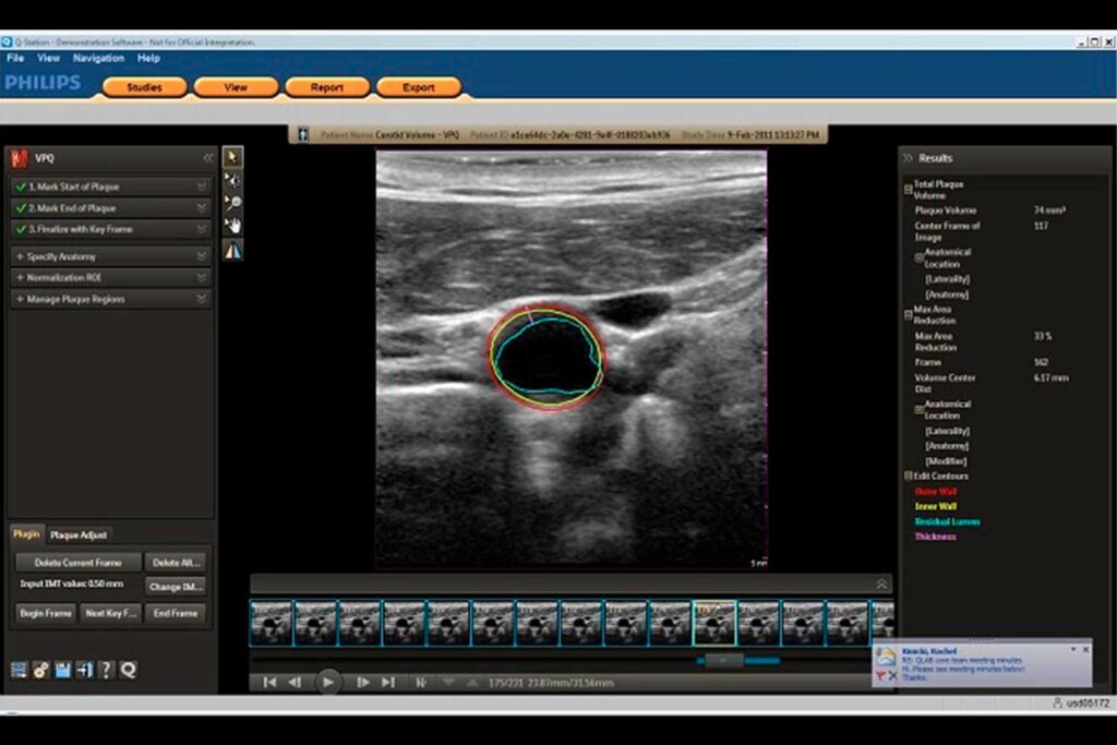
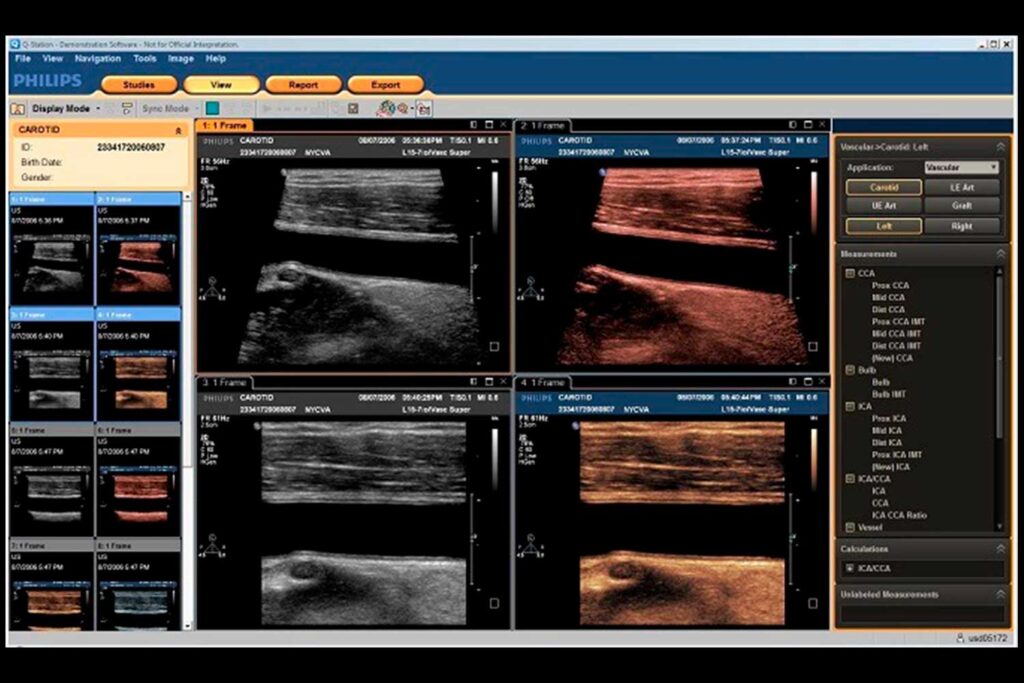
The built-in, customizable, and extensible Q-App includes comprehensive analysis and quantification capabilities. The app allows you to view echo studies, analyze and quantify 2D/3D data, save screenshots in DICOM format, view and edit measurements, include conclusions and comments in reports, and save results.
Advanced 3D visualization
Integrated QLAB applications allow easy viewing of 3D echo data. You can view 3D data sets, including mitral valve images, from any arbitrary point and in any arbitrary projection. Using the Q-Station application, you can save processed and reconstructed images in the patient study and export them to the PACS station. Simple and reproducible quantitative analysis of cardiac structures, analysis of data obtained with contrast, evaluation of breast tissue, a semi-automated protocol for evaluation of the fetal heart – these are just some of the capabilities available in the Q-Station environment.
Cardiology
Dedicated quantitative analysis of cardiac mechanics under stress with QLAB CMQ Stress and 2D speckle tracking. Stress protocols automatically link wall motion assessment to test stages and anatomical views – a single click changes both stage and view, ensuring continuous synchrony of data acquisition. Saving preferred loop sections from EPIQ and iE33 systems takes the operator to a new level of stress echo workflow that improves productivity. Step-by-step user interface and controls allow you to customize your scan protocol. Learn quickly and integrate easily into your workflow.
Q-Station application – the core of the workflow
Manage studies, series, images and reports. Recover data from local databases, CDs, DVDs, USB sticks and PACS stations. Copy, move and merge studies, forward preliminary ultrasound protocols by email.
QLAB package capabilities enable specialized analysis and quantification
You can obtain an ejection fraction value in less than a minute to objectively assess overall left ventricular function. Automated region of interest tracking saves time and improves reproducibility. Comprehensive 2D image analysis, viewing 3D data sets from any arbitrary point and in any arbitrary projection, and mitral valve assessment. All analyses and extracted data are easily incorporated into ultrasound protocols.
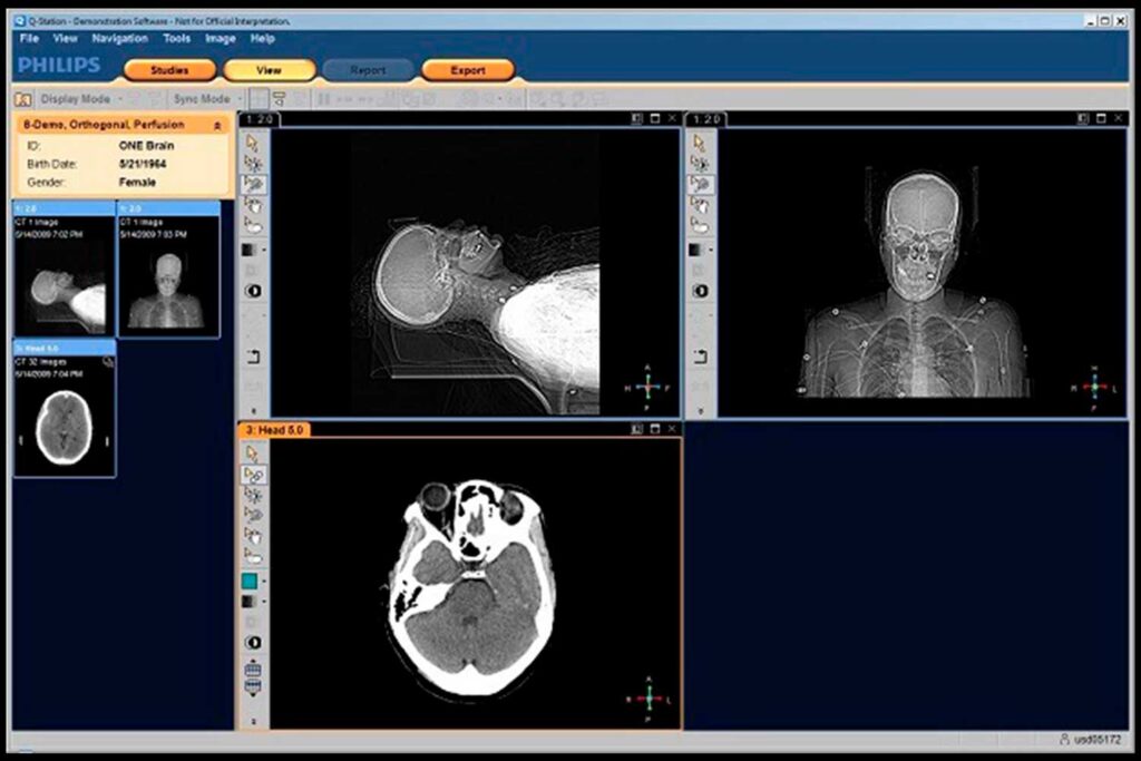
Viewing non-ultrasound images: CT, MRI, X-ray angiography, NM and
Viewing non-ultrasound control images provides a broader understanding of the patient’s condition. Viewing ultrasound images and images obtained by other methods on one screen allows you to visually monitor the effectiveness of the patient’s treatment at all stages.