

 РУ
EP navigator
Catheter navigation tool for EP procedures
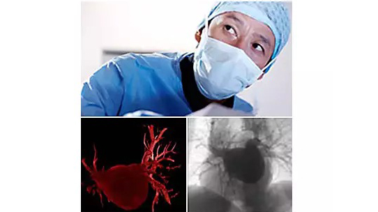
EP navigator facilitates intuitive 3D catheter image guidance during AF ablation procedures. It provides a detailed 3D image of anatomy, which can be precisely registered and overlaid onto 2D live fluoroscopy to support complex procedures.
Description
Automatic segmentation
The 3D volume is automatically segmented to show the left atrium and pulmonary veins. During segmentation, you can measure the pulmonary veins (PV) and left atrium dimensions (LA).
Export to mapping
EP navigator precisely registers the 3D volume with the live 2D fluoroscopy images. The segmented volume can also be exported to standard mapping systems to manage dose, mapping time, and the need to re-registerdata.
View different anatomical planes
During Live Image Guidance, you can view the rotational scan by different anatomical planes to get an immediate reference point. The 3D volume can also be displayed with an outline around critical structures to support decision making.
EP navigator and reduced 3D rotational scan
EP navigator offers the option of reduced angular 3D rotational scanning on the Allura X-ray system. You can create an excellent 3D image of cardiac structures at the precise time of the procedure* to plan the optimal therapeutic approach.