

 РУ
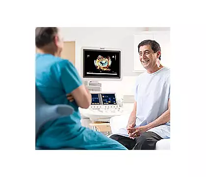
Live 3D TEE
3D Ultrasound
With Live 3D transesophageal echocardiography, cardiologists, cardiac surgeons, anesthesiologists, interventional cardiologists and echocardiologists can observe the structure and function of the heart in real time. This method is fast, reproducible and can be quantitatively analyzed.
Contact usDescription
Structure and Function
The ability to observe the structure and function of the heart in real time, even in difficult-to-scan cases.
Visualization
Visualization for device implantation
Leadership in Live 3D Transesophageal Echocardiography
Philips xMATRIX Live 3D Transesophageal Echocardiography was introduced in 2007 and has since been successfully used in millions of exams and procedures worldwide.
Data and projections
Access to data and projections not accessible transthoracically.
Improved image quality
Improved image quality during ultrasound-guided procedures
Monitoring
Monitoring before, during and after surgery.